AI DERMATOLOGY
Fast, reliable data
Delivering specialist-level1 data to help inform diagnosis and treatment selection
- Accurately identifies inflammatory cells and differentiates between cocci and rods1
- Reliable performance comparable to ACVP clinical pathologists
- Consistent point-of-care identification of inflammatory cells, bacteria and yeast
Study Design
Vetscan Imagyst® AI Dermatology was evaluated in 143 dog and 75 cat samples and compared to that of board-certified clinical pathologists.1
- Samples included skin impression smears, ear swabs and skin swabs
- Each sample was classified as positive or negative for inflammatory cells or infectious agents by the consensus of 2 of 3 clinical pathologists
Inflammatory cell detection with expert-level accuracy1
| MACROPHAGES | EOSINOPHILS | LYMPHOCYTES | NEUTROPHILS* | |
|---|---|---|---|---|
| Sensitivity (95% CI) |
96% (91%-99%) |
93% (83%-97%) |
86% (79%-91%) |
95% (90%-98%) |
| Specificity (95% CI) |
84% (76%-90%) |
94% (89%-97%) |
85% (77%-91%) |
95% (89%-98%) |
95% Jeffreys confidence interval
Highly precise infectious agent detection and accurate bacteria differentiation1
- 89% positive predictive value for identifying cocci- and rod-shaped bacteria†
| COCCI BACTERIA | ROD BACTERIA | MALASSEZZIA1 | |
|---|---|---|---|
| Sensitivity (95% CI) |
86% (80%-92%) |
83% (72%-91%) |
92% (82%-97%) |
| Specificity (95% CI) |
82% (73%-89%) |
76% (69%-82%) |
87% (81%-91%) |
95% Jeffreys confidence interval
The microscopic findings from a cytological evaluation should always be interpreted considering the clinical findings.2
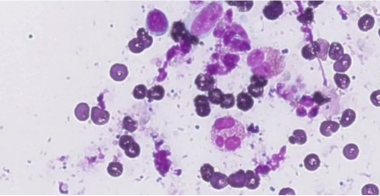
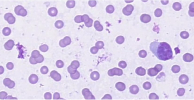
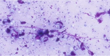
High-resolution images for powerful insights
The AI Dermatology algorithm performance in the evaluation of the scan area was comparable to that of expert clinical pathologists.1
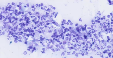
Macrophages
Eosinophils
Lymphocytes
Neutrophils containing bacteria
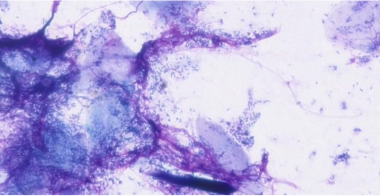
Rod-shaped bacteria
.png)
Cocci-shaped bacteria
-(2).png)
Malassezia

Bringing specialist-level medicine to your clinic
The Zoetis Virtual Laboratory is an integrated support network of board-certified specialists paired with expert-level AI1,3-9, providing actionable insights to help you diagnose and treat with confidence.
- Best-in-class AI
- Anytime‡ expert support
- Connected diagnostic insights
Discover the difference Vetscan Imagyst can make in your clinic
Contact us today to learn more about how our diagnostics portfolio can help you provide elevated patient care.
Customer Service, Support & Ordering
Mon – Fri: 8:30am – 6:30pm ET
Vetscan and i-STAT Technical Support
24 hours a day, 7 days a week, 365 days a year
1-888-963-8471
Zoetis Reference Laboratories Customer Service, Support & Ordering
Mon – Fri: 8:00am – 9:30pm ET
Saturday: 9:00am – 8:30pm ET
Sunday: Closed
1-888-965-9652
Need Technical Support?
Our support team is available to answer product questions and provide guidance.§
* Including Malassezia on keratinocytes.
† Defined as the number of true positives divided by true positives plus false positives.
‡ Dependent on consultant availability.
§ If you are a pet owner looking for treatment recommendations, please contact your veterinarian.
References: 1. Data on file, Study No. DHX6Z-US-23-222, 2023, Zoetis Inc. 2. Fisher D. Cutaneous and subcutaneous lesions. In: Cowell RL, Valenciano AC. Cowell and Tyler’s Diagnostic Cytology and Hematology of the Dog and Cat. 5th ed. Elsevier; 2019:74-101. 3. Data on file, Study No. DHX6Z-US-23-205, 2024, Zoetis Inc. 4. Data on file, Study No. DHX6Z-US-23-206, 2024, Zoetis Inc. 5. Data on file, Study No. DHX6Z-US-23-209, 2024, Zoetis Inc. 6. Nagamori Y, Hall-Sedlak R, Blagburn B, et al. Multicenter evaluation of the Vetscan Imagyst system using Ocus 40 and EasyScan One scanners to detect gastrointestinal parasites of feces in dogs and cats. Journal of Veterinary Diagnostic Investigation. 2023;36(1). doi: 10.1177/10406387231216185. 7. Data on file, Study No. DHXMZ-US-23-218, 2023, Zoetis Inc. 8. Data on file, Study No. DHX6Z-US-22-131, 2022, Zoetis Inc. 9. Data on file. Study No. DHXMZ-US-24-235, 2024, Zoetis Inc.